(Please click on blue terms for more information and purple links for further explanation from external websites)
It is unfortunate that women are far more likely to have problems with their bodies.
For example, whilst 1 in 8 women in the UK are diagnosed with breast cancer annually there are on average only 300 cases of male breast cancer diagnosed annually.
Although men can have cancer of the testicles and prostate gland, women can have cancer of the womb, ovaries and cervix.
Where State-of-the-Art systems are available breast and other cancers affecting women are being treated without surgery using High Intensity Focused Ultrasound.
In addition to cancer, there are other types of growths and conditions that can affect the womb/uterus, which are also being treated without surgery using this system, e.g. Uterine Fibroids.
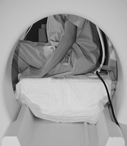
The video shows how Uterine Fibroids are treated with High Intensity Focused Ultrasound. (An MRI scanner provides the imaging for the treatment). This system can also be used to treat breast cancer and other cancers, and other conditions such as
Endometriosis, Adenomyosis.
Click Image for Video
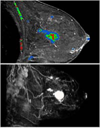
The following images of breast cancer have been taken using MRI. (The 3D colour enhancement is provided by advanced computer technology). These images show the location and extent of the cancer and are provided on screen during treatment, enabling complete removal.
(State-of-the-Art systems have been successfully used for over 20 years, primarily in China but also in other countries such as Japan, USA, Russia, for the imaging and treatment of conditions affecting women, e.g. breast and womb/uterine cancer and other conditions such as Uterine Fibroids, Endometriosis and Adenomyosis).
Importantly, there is no need for surgery - for removal of part or all of the breast, womb or ovaries - for any condition affecting them that can be treated with High Intensity Focused Ultrasound.
The conditions of Uterine Fibroids, Endometriosis and Adenomyosis can not only cause much pain and problems with becoming pregnant but can also affect the normal functioning of abdominal organs, e.g. the intestine, bowel and bladder.
Apart from these conditions pregnancy, childbirth and intervention in childbirth such as episiotomies, forceps delivery, and surgery, e.g. caesarean section, can also affect the normal functioning of these organs and others such as the rectum and anus.
Some of the problems which can result are, difficult evacuation, bloating, constipation, blockages (obstruction), restriction, inflammation, inability to pass urine, urinary incontinence, faecal incontinence, having abdominal and/or rectal pain, and pain during intercourse.
Some further images and information of the benefit of State-of-the-Art systems for the diagnosis and treatment of these conditions follow the diagram.
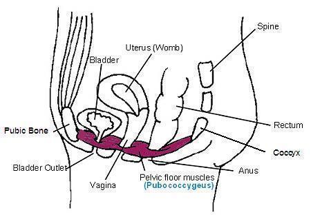
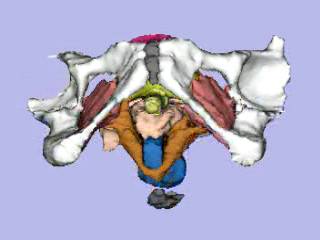
Below is a typical diagram of the ‘muscles of the pelvic floor’ (lack of support/weakness in which is often attributed to problems with normal functioning. However, a flat (one dimensional) image can really only show a muscle called the
pubococcygeus).
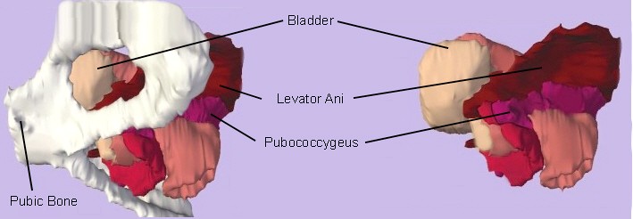
Side View of Body
The following is a 3D colour MRI image of the ‘muscles of the pelvic floor’ taken from a side view of the body. This image shows the pubococcygeus is attached to other muscles (including a larger and more important muscle in support and functioning called the levator ani).
Note: For anyone who does not have medical training it is possible that the above and following 3D colour images and movies are not easily understood, but those who have produced them and the clinicians who use them, say they are vital for comprehensive and accurate diagnosis and treatment and that surgery should not be undertaken without them.
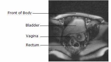
An example MRI image taken on the most advanced UK system for the diagnosis of functioning problems relating to the female abdomen/pelvis is below, e.g. the cause of urinary incontinence. This image has been taken looking upwards through the body and is one of many which have been viewed by an International Expert on MRI, his comments are, "It is only possible to form an impression of the cause of problems with these images and they would be much easier to read in movie format."
The following 3D colour MRI image of the female abdomen/pelvis (from an overseas treatment centre) can be shown in movie format which allows the bones, organs, muscles to be seen individually and from all angles. Importantly, these images show the Levator Ani muscle (brown) is attached to the pelvis differently on the right side to the left, and this difference in attachment was the cause of urinary incontinence.
Colours in Image
bones
white
coccyx and pubis
grey
levator ani muscle
brown
bladder
yellow
rectum
blue
vagina
pink
uterus/womb
cerise
Click image for Movie
Without imaging, the cause of urinary incontinence might for example be attributed to a condition known as a Cystocele, and any surgery to correct a Cystocele would therefore not remedy the problem.
3D colour images and movies can be provided on screen before, during and after procedures.
This advanced imaging enables precise treatment as a patient's body and problems with it are shown comprehensively and accurately throughout procedures and statistically this has shown to improve treatments and outcomes.
Conditions such as rectoceles, enteroceles, hernias, and others can also be identified and treated using these systems. As all of these conditions can affect the function of the intestine and bowel which can then cause other problems, it is important that imaging can identify them.
The following is another type of 3D MRI, the movie shows a rectocele (Note: movies which show movement are also called Dynamic).
This is from a diagnostic and treatment centre in the States (the bold text is theirs). It shows a side view of the pelvis.
Dynamic MRI of the pelvic organs is a modality for imaging the pelvis. It helps doctors get a clearer picture of the problem, so that it can be better addressed. The dynamic pelvic MRI below demonstrated a rectocele (outward bulging in the rectum). Unlike normal 'X rays', the images play like a short movie. It shows a large protrusion in the lower rectum, and it gives a more complete picture because the other pelvic organs are visualized.
Click image for Movie
As said previously, these images may not be easily understood but overseas clinicians say no surgery should be undertaken without them as they show the cause of problems and without imaging problems can be made worse. They also say movie images of the pelvis should be done in the way a patient experiences problems, e.g. seated and whilst applying abdominal pressure. In the UK there are no facilities to provide this.
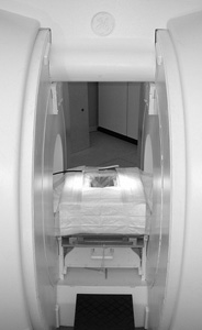
An example of an MRI scanner which can provide imaging to show the cause of pelvic dysfunction is below. (The ‘toilet’ can be removed, so the patient can assume various positions in the scanner, e.g. lie on their back and because there is ample room bend their knees and bear down as in childbirth, to show any problems caused by this).
MRI Side View
MRI Front View
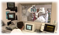
Any surgical correction required can be carried out in the scanner due to the evident access and procedures can be shown on screen throughout, called real-time image guided surgery/treatment.
As regards real-time image guided surgery/treatment this is said to be essential for patient well-being, some images of operating rooms follow. All enable a radiologist (one who reads scans) to assist a surgeon throughout procedures and also enable a surgeon to view a patients body on screen throughout procedures.
Reported operational advantages: 3D imaging while surgery is undertaken allows for multiple teams to manage complicated conditions which results in better outcomes - patients problems will be identified and confirmed through precise images - increased success rate of "first attempt" surgeries.
Some further information from overseas clinicians:
Magnetic resonance imaging (MRI) is essential for the diagnosis of pelvic floor dysfunction, given the inaccuracy of clinical examination which fails in near to 90% of patients. It is a major health problem affecting up to 50% of women with approximately 30% of women who undergo surgical repair requiring another surgery for recurrence of symptoms.
With many thanks to the dedicated and gifted physicians, radiologists, computer scientists, software developers, engineers, and other researchers due to whose untiring efforts these technologically advanced systems are available. Also with many thanks to Rob Key for his efficient help and expertise - contact enquiries@latchkeydesign.co.uk for website design and alterations.
We close with views from the medical profession on some of the values of these amazing technologies, ‘3D imaging allows us to see the problem’, ‘mastectomies (removal of the breast) and hysterectomies (removal of the womb) will probably become obsolete as forms of treatment’. ‘Imaging is the gateway to new non-invasive treatments’.
Please help to make the benefits of State-of-the-Art systems available for women in the UK. (Your support could help so many so please click here to Donate or Fundraise)


/Resources/Dynamic Pelvic MRI showing rectocelesmall.jpg)